
Leg Ulcer Wound Care
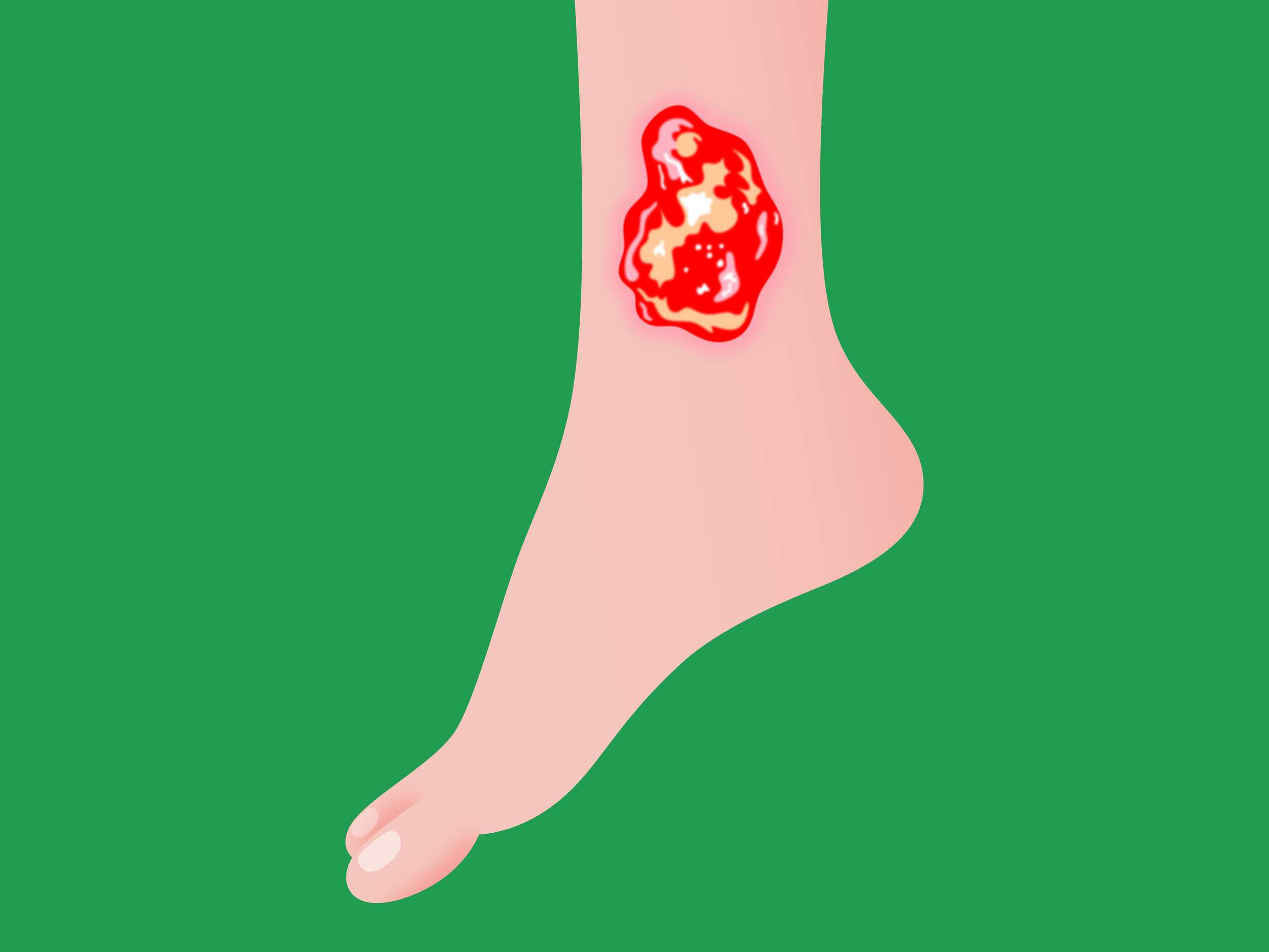
Leg ulcers are persistent, long-lasting sores that take longer than 4 to 6 weeks to heal. Usually, they start to form slightly above the ankle on the inside of the leg. In addition to discomfort, swelling, and itching in the affected leg, venous leg ulcers can also cause anemia.
Why do I have Leg Ulcers?
Acute wounds heal under normal circumstances. Wounds fail to heal and become chronic when healing is physiologically compromised. The necessary components for proper healing are the following:
Good vascularization.
Absence of necrotic tissue.
Infection-free.
Remaining moist.
Causes of Leg Ulcers
Diabetes complicated by peripheral vascular disease (PVD) (arterial and venous) and neuropathy.
Non-diabetic peripheral arterial disease.
Non-diabetic chronic venous disease.
Non-diabetic peripheral neuropathy.
Smoking, which introduces vasoconstriction in the process and other atherosclerotic conditions such as hypertension only augment the ongoing devitalization of tissue. Trauma can be an external cause of leg ulcers.
Less common causes include infection, vasculitis, malignancy, drugs, and spider bites.
The foundation for all healthy tissue is a good blood supply. Diabetes and peripheral vascular disease impact vital tissues and nerves negatively due to compromise in blood supply and the resulting hypoperfusion, with the hypoxia, neuropathy, and necrosis of tissue that follow.
Tissue breakdown manifests as ulceration.
Venous insufficiency is the most common cause of leg ulcers. Atherosclerosis affects the arteries with limited blood flow through them.
Venous Insufficiency-Related Leg Ulcers
Venous insufficiency rises with age, obesity, gender (F > M), pregnancy, prolonged standing (gravity), and a history of deep vein thrombosis (DVT).
Clinical examination is sufficient to make the diagnosis. Spider veins (telangectasias) of the feet and ankles, edema, varicose veins, and brown discoloration (hemosiderin deposits) are common findings. Location is typically from mid-calf to ankle. There is hemosiderin deposition in macrophages that darken the surrounding skin. Stasis dermatitis is inflammatory changes due to venous backup. Edema is common.
Diagnosis is confirmed via ultrasound.
Arterial Disease-Related Leg Ulcers
These are caused by cigarette smoking, diabetes, abnormal lipids, and arterial obstructions. They typically occur at distal points, such as the toes, and at pressure sites overlying bony prominences. There is usually hair loss and the atrophic skin takes on a shiny appearance.
Diagnosis is confirmed via computed tomography (CT) angiography and/or magnetic resonance imaging (MRI) angiography.
Neuropathic Ulcers
These present as “punched-out” ulcers over pressure points on the foot or heel most commonly. There is usually callus formation around them. Reduction of sweating (hypo- or anhidrosis) can cause the feet to be dry and scaly.
The diagnosis of ulceration in the legs must include consideration for its cause so that both the ulcer and the tendency to progress can be addressed. As such, tests for the following are indicated:
Diabetes testing: Including screening tests (2 hour glucose tolerance test after a 75 g glucose load orally).
For established diabetics: Monthly measures of the glycated hemoglobin A1C.
Evaluation of the blood supply to the lower extremities to rule out arterial vascular disease or chronic venous disease: arteriography imaging, ultrasound.
Evaluation of the peripheral nervous system: For sensory perception as well as chronic vasoconstriction influences from the autonomic nervous system.
Testing for the causes of hypertension, cardiovascular disease (CVD), and atherosclerosis: Including lipid panels to identify dyslipidemia (abnormal cholesterol and triglycerides).
How do I manage and treat my Leg Ulcer?
Optimal wound healing requires well-vascularized tissue, absence of necrotic tissue, infection-free conditions, and moisture.
Dressing Leg Ulcers
Dressing should be applied to the wound with even distributive contact, and should control exudate, hamper bacterial growth, and be easy to manage for both healthcare professionals and the patient. At a point at which wounds demonstrate progressive healing, consideration for closure is possible.
Debridement involves cutting away dead (vitalized) tissue. Topical antiseptics and antimicrobials can be used, but with caution due to their risk of causing local toxicity.
For very deep wounds, negative pressure dressing is used which utilizes a suction mechanism to allow the tissues to collapse and reduce the depth of the defect.
Granulation tissue is new growth and fill of well-vascularized tissue and signifies successful management with the opportunity for closure.
Other exotic treatments include hyperbaric oxygen therapy (HBOT) and electrical stimulation, with mixed results.
How can I prevent Leg Ulcers?
Prevention of leg ulcers requires both on-going treatment for present ulcerations and preventive measures against future ulcerations.
On-going treatment includes antimicrobials and dressing strategies with close surveillance of ulcerations.
Prevention of treatment failure and future ulcers relies on:
Smoking cessation: The process of discontinuing tobacco use.
Good nutrition: With the aid of consultation with a dietitian/nutritionist.
Mobilization: Prolonged immobilization increase the risk of treatment failure and for new ulcerations. If self-ambulation or mobilization is impossible due to handicaps, passive motion and physical therapy can be used.
Avoidance of immunosuppressive drugs: Inflammation is only one stage of the continuum of healing, and suppressing it can have more risk than benefit in the healing process.
Diabetics: Strict glycemic control.
Management of peripheral vascular disease (PVD): Ischemia my require re-vascularization procedures. Chronic venous insufficiency can be addressed via compression stockings, weight loss, ambulation, and avoidance of prolonged sitting or standing.